八月 26, 2021
1978
The technological advances achieved in the field of medical imaging in the last century have created unprecedented opportunities for non-invasive diagnosis and established medical imaging as an integral part of the medical health system. One of the main areas of innovation that represents these advances is the interdisciplinary field of medical image processing.
This rapidly developing field involves a wide range of processes from raw data acquisition to digital image transmission, and these processes are the basis for the complete data flow in modern medical imaging systems. Nowadays, these systems provide higher and higher resolutions in spatial and intensity dimensions, as well as faster acquisition times, resulting in a large amount of high-quality raw image data, which must be processed and interpreted correctly to obtain accurate diagnostic results.
Core areas of medical image processing
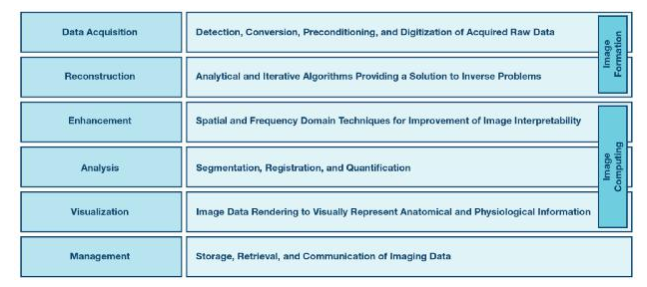
There are many concepts and methods used to construct the field of medical image processing. These concepts and methods focus on different aspects of its core area, as shown in Figure 1. These aspects form the three main processes in this field-image formation, image calculation and image management.

The image formation process consists of data acquisition and image reconstruction steps, used to solve mathematical inversion problems. The purpose of image calculation is to improve the interpretability of reconstructed images and extract clinically relevant information from them. Finally, image management deals with the compression, archiving, retrieval and transmission of acquired images and derivative information.
image formation
data collection
The first necessary step of image formation is to collect raw imaging data. This data contains raw information about the captured physical quantities that describe the internal organs of the body. This information becomes the main subject of all subsequent image processing steps.
"Different types of imaging modes can use different physical principles, which involve the detection of different physical quantities. For example, in digital radiography (DR) or computer tomography (CT), it is the energy of the incident photon; in positron emission tomography (PET), it is the photon energy and its detection time; in magnetic resonance imaging ( In MRI), it is the parameter of the radio frequency signal emitted by the excited atoms; in the ultrasound, it is the echo parameter.
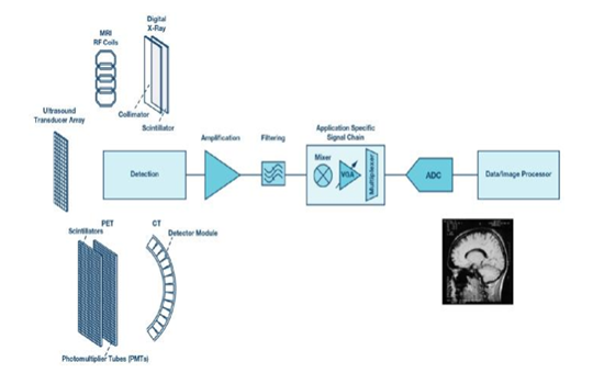
However, regardless of the type of imaging mode, the data acquisition process can be subdivided into the detection of physical quantities, including the conversion of physical quantities into electrical signals, pre-conditioning of the collected signals, and the digitization of physical quantities. A general block diagram showing that all these steps are applicable to most medical imaging modes is shown in Figure 2.

Image reconstruction
Image reconstruction is the mathematical process of forming an image using the acquired raw data. For multi-dimensional imaging, the process also includes the combination of multiple data sets captured at different angles or at different time steps. This part of medical image processing solves the problem of inversion, which is the basic theme of this field. There are two main types of algorithms used to solve such problems-analysis and iteration.
Typical examples of analytical methods include filtered back projection (FBP), which is widely used in tomography; Fourier transform (FT), which is particularly important in MRI; and time-delay superposition (DAS) beamforming, which is a type of ultrasound examination. Indispensable technology. These algorithms are sophisticated and efficient in terms of required processing power and computing time.
However, they are based on idealized models and therefore have some obvious limitations, including their inability to deal with complex factors such as the statistical properties of measurement noise and the physics of the imaging system.
The iterative algorithm overcomes these limitations and greatly improves the insensitivity to noise and the ability to reconstruct the optimal image using incomplete original data. Iterative methods usually use systematic and statistical noise models, and calculate projections based on the initial target model using assumption coefficients. The difference between the calculated projection and the original data defines a new coefficient used to update the object model. This process is repeated using multiple iterative steps until the cost function of the mapping estimated value and the true value is minimized, thereby integrating the reconstruction process into the final image.
There are many kinds of iterative methods, including maximum likelihood expectation maximization (MLEM), maximum a posteriori (MAP), algebraic reconstruction (ARC) technology, and many other methods currently widely used in medical imaging modes.
Image calculation
Image calculation involves calculations and mathematical methods for computing reconstructed imaging data, which are used to extract clinically relevant information. These methods are used for enhancement, analysis and visualization of imaging results.
Enhance
Image enhancement optimizes the transformation of the image to improve the interpretability of the information contained. The method can be subdivided into spatial domain and frequency domain technology.
spatial domain technology directly acts on image pixels, which is particularly useful for contrast optimization. These techniques usually rely on logarithms, histograms, and power law transformations. The frequency domain method uses frequency transformation and is most suitable for smoothing and sharpening images by applying different types of filters.
Using all these technologies can reduce noise and unevenness, optimize contrast, enhance edges, eliminate artifacts, and improve other related features that are critical to subsequent image analysis and accurate interpretation.
analyze
Image analysis is the core process in image calculation. The various methods it uses can be divided into three categories: image segmentation, image registration, and image quantization.
The image segmentation process divides the image into meaningful contours of different anatomical structures. Image registration can ensure the correct alignment of multiple images, which is particularly important for analyzing time changes or combining images acquired using different modes. The quantification process determines the properties of the identified structure, such as volume, diameter, composition, and other relevant anatomical or physiological information. All these processes directly affect the inspection quality of imaging data and the accuracy of medical results.
Visualization
The visualization process presents image data as an intuitive representation of anatomical and physiological imaging information in a specific form in a defined dimension. By directly interacting with the data, visualization can be performed in the initial and intermediate stages of imaging analysis (for example, to assist in the segmentation and registration process), and the optimized results can be displayed in the final stage.
Image management
The last part of medical image processing involves the management of acquired information, including various technologies for image data storage, retrieval, and transmission. Several standards and technologies have been developed to handle all aspects of image management. For example, the medical imaging technology Image Archiving and Transmission System (PACS) provides economical storage and access to images from multiple modes, while the Digital Imaging and Communication in Medicine (DICOM) standard is used to store and transmit medical images. The special technology of image compression and streaming achieves these tasks efficiently.
Challenges and trends
Medical imaging is a relatively conservative field, and it usually takes more than ten years to transition from research to clinical application. However, its nature is complex, and it faces many challenges in all aspects of its constituent scientific disciplines, which steadily promotes the continuous development of new methods. These developments represent the main trends that can be determined in today's core field of medical image processing.
The field of image acquisition benefits from innovative hardware technologies developed to improve the quality of original data and enrich its information content. The integrated front-end solution can achieve faster scan time, finer resolution and advanced architecture, such as ultrasound/mammography, CT/PET or PET/MRI combined systems.
Fast and efficient iterative algorithms have replaced analysis methods and are increasingly used for image reconstruction. They can significantly improve the image quality of PET, reduce the X-ray dose in CT, and perform compression detection in MRI. Data-driven signal models are replacing artificially defined models, providing better solutions to inversion problems based on limited or noisy data. The main research areas that represent the trends and challenges of image reconstruction include system physical modeling and signal model development, optimization algorithms, and image quality assessment methods.
As imaging hardware captures more and more data, algorithms become more and more complex, and people urgently need more efficient computing technology. This huge challenge can be solved by a more powerful graphics processor and multi-processing technology, providing new opportunities for the transition from research to application.
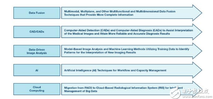
The main trends and challenges related to this transformation of image computing and image management cover many topics, some of which are shown in Figure 3.

The continuous development of new technologies related to all these topics has narrowed the gap between research and clinical applications, promoted the integration of medical image processing and physician workflows, and ensured more accurate and reliable imaging results.
ADI provides a variety of solutions to meet the most demanding medical imaging requirements for data acquisition electronic design, including dynamic range, resolution, accuracy, linearity and noise. Below are a few examples of such solutions developed to ensure the highest initial quality of raw imaging data.
The highly integrated analog front end ADAS1256 with 256 channels is specially designed for DR applications. The ADAS1135 and ADAS1134 multi-channel data acquisition systems with excellent linearity can maximize the image quality of CT applications. The multi-channel ADC AD9228, AD9637, AD9219, and AD9212 are optimized for excellent dynamic performance and low power consumption, which can meet PET requirements. The pipeline ADC AD9656 provides excellent dynamic performance and low power consumption for MRI. The AD9671 integrated receiver front end is designed for low-cost, low-power medical ultrasound applications that require a small footprint.
in conclusion
Medical image processing is a very complex interdisciplinary field, covering many scientific disciplines from mathematics and computer science to physics and medicine. This article attempts to propose a simplified but well-structured core domain framework that represents the domain and its main themes, trends, and challenges. Among them, the data acquisition process is the first and one of the most important areas, which defines the initial quality level of the raw data used in all subsequent stages of the medical image processing framework.